Transmission electron microscopy (TEM) laboratory
Applications of EM for analysis of biological samples at the EM lab in IMCB
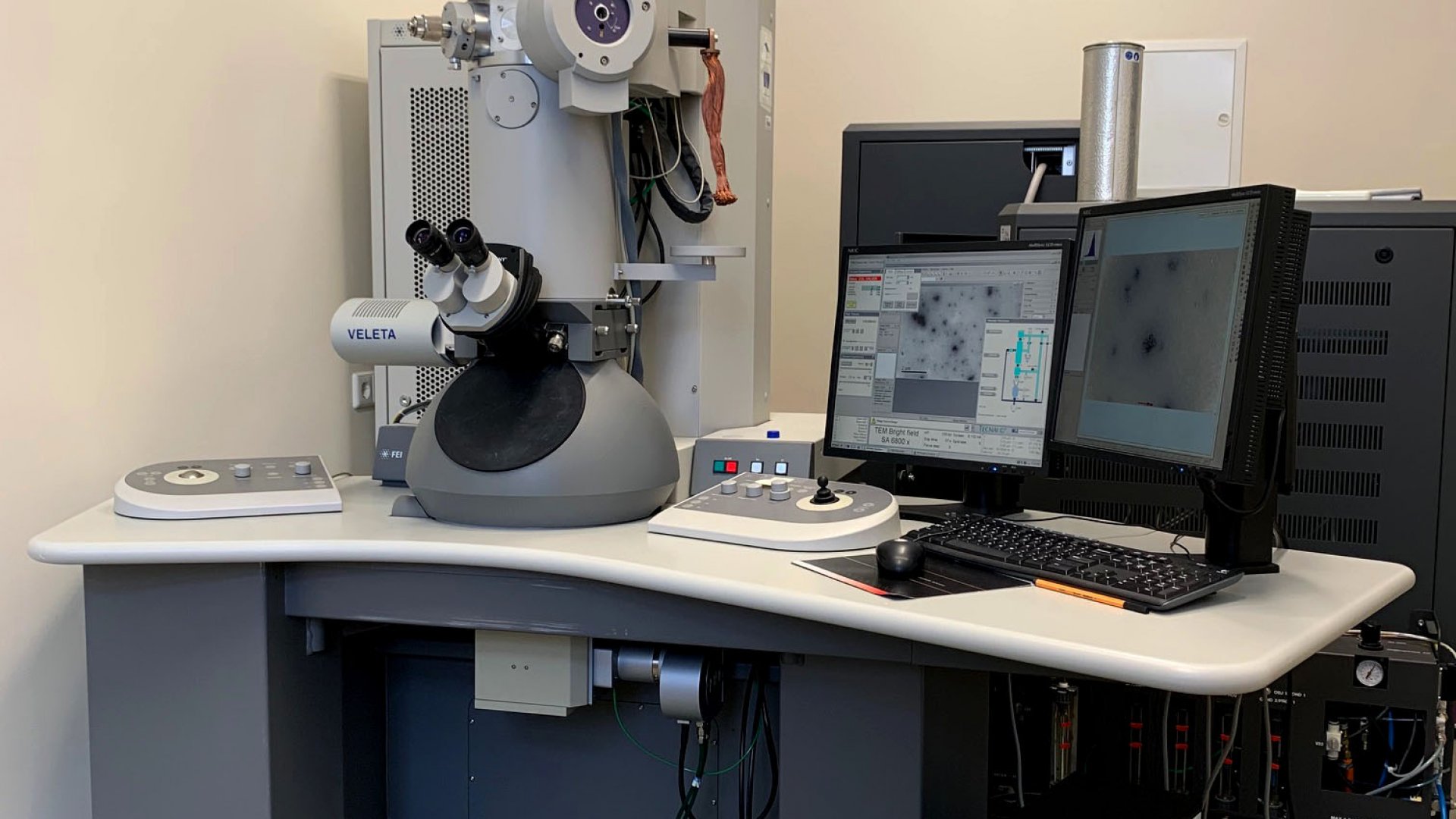
The FEI Tecnai G2 Spirit BioTwin allows users to perform microscopy for life sciences applications. The lens configuration guarantees maximum contrast and the TEM is optimized to provide high-resolution imaging and analysis with conventional thin-sectioned biological specimens and routine negative stained samples..
Equipment technical specifications (PDF)
Equipment:
Transmission electron microscope
Model: Tecnai G2 Spirit BioTWIN (Philips Electron Optics, Holland)
Location: Vanemuise Street 46, room 135
Resolution (nm):
• Line: 0,344
• Point: <0,490
Electron source: volfram-elektrood või LaB6
Voltage: 20 - 120 kV
Magnification: 22 – 340 000 x
Equipped with:
• Gatan Orius SC 1000 bottom mounted CCD camera, resolution 11 megapixel (4008 x 2672 pixels) (Gatan Inc., USA), (for technical details see Orius brochure)
• Olympus-SIS Veleta side-mounted CCD camera,resolution 2048 x 2048 pixels) (Olympus SIS GmbH), (for technical details see Veleta brochure)
• FEI CompuStage single-tilt standard specimen holder
The FEI specimen holder is delicate and fragile as it is designed for high-tilt ( +/- 70 deg) computer controlled goniometers. The microscope is equipped with fully computer-controlled, eucentric side-entry, high-stability CompuStage, which enables the specimen to move at all XYZ positions.
Software:
• Digital Micrograph software for operating the Gatan camera
• FEI TEM Imaging & Analysis (TIA) software for recording and processing images from both Veleta and Gatan cameras
Ultramicrotomes
The ultramicrotomes are mainly used to prepare thin sections for imaging with TEM. Either glass or diamond knifes can be used to cut thin sections (60-100 nm as standards) of specimens embedded in epoxy resin.
Model: Leica Ultracut EM UC7 (Leica Microsystems GmbH, Austria)
Section thickness: up to 15 µm
Cutting window: 0,2 mm up to 14 mm adjustable
Cutting speed: 0,05 – 100 mm/s
Knife holder: 360° rotatable
Specimen holder: 360° rotatable
Equipped with:
• Leica EM FC7 cryochamber, low temperatuure sectioning system for the Leica UC7 ultramicrotome. It is used for semi- and ultrathin cryosectioning of biological samples such as frozen hydrated sectioning of vitrified specimens and cryoprotected biological samples in Tokuyasu technique at the temperatuure of -15 °C to -185 °C.
• Leica EM CRION, antistatic device for working with cryochamber.
Model: RMC PowerTome MT-XL (Boeckeler Instruments, Inc., USA)
Section thickness: 5 nm to ~10 µm
Cutting window: adjustable from 0,2 mm to 15
Cutting speed: 0,1 – 49,9 mm/s (adjustable by 0,1 mm/s step)
Knife holder: 360° rotatatable
Specimen holder: 360° rotatatable
Model: Reichert Jung 2050 Supercut (produced by C. Reichert Optische Werke AG in Vienna, Austria, between 1950-1990. The former Reichert-Jung company is now called Leica Microsystems GmbH)
Section thickness: ~2 -15 µm
The Reichert Jung 2050 SuperCut Motorized Microtome is the rotary microtome for sectioning mainly the delicate semi-thin plastic sections.
Vacuum-coater
Vacuum coater is a device for precise coating of samples with carbon layer for subsequent examination with an electron microscope. Carbon coating is achieved by carbon thread evaporation. The coater is equipped with integrated quartz crystal film thickness measurement, which accurately determines the layer thickness. The vacuum-coater is also suitable for the glow discharge process which is used to make TEM grids hydrophilic.
Model: Leica EM ACE600 (Leica Microsystems GmbH, Austria)
Ultimate vacuum: ≤ 2x10⁻⁶ mbar
Coating time: 1 – 1800 sec
Coating thickness: 1 – 1000 nm
Quartz crystal measurement resolution: one decimal (0.1 nm accuracy)
Glow discharge: 5 – 15 mA, continously adjustable
Pictures from IMCB TEM laboratory
The pictures show the devices described above.


